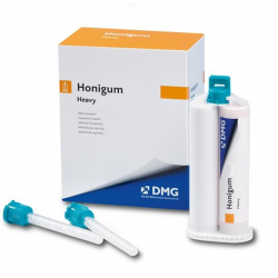
The erosion infiltration technique, a minimally invasive approach of management of enamel white spots, was proposed and discussed with the consent of the patient. After the clinical and radiographic examination, the occurrence of hypomineralization of the upper first molars characterizing MIH confirmed the diagnosis.
Mini dam dmg free#
The periodontal status of the patient was healthy and free from visible plaque, and the radiographic examination revealed no abnormalities of the supporting tissues. The spots were well limited, with distinct borders from the adjacent enamel (Figures 1(a) and 1(b)). The spots were easily noticeable in the frontal view of the anterior teeth: big chalky white spots on tooth number 21 and 11 and two less pronounced spots on tooth 12 and 22. Intraoral examination revealed white spots in the upper bilateral central and lateral incisors. The anamnesis did not specify any systemic illnesses of the patient. Case PresentationĪ 29-year-old woman was referred to the Department of Dental Medicine of Hospital of Charles Nicolle, Tunis, with the chief complaint of restoring her smile before her wedding soon. This paper is aimed at describing and illustrating the aesthetic management of white opacities caused by MIH using a resin infiltration technique. Recently, a resin infiltration technique was introduced with the development of highly flowable resin material. The common treatment strategy comprises restorative procedures, improvement of remineralization using CCP-ACP-containing or fluoride-containing products, microabrasion, and bleaching technique laminate veneer restoration. Several techniques have been proposed to improve the appearance of MIH spot lesions. Incisal enamel defects are frequently quite extensive and most common on the buccal surfaces of the teeth, giving rise to more esthetic concerns. The opacities range from white to yellow/brown in appearance, and in lesions with a severe mineral deficit, rapid progression to posteruptive enamel breakdown can occur. The defining clinical features of MIH are demarcated opacities with clear, distinct borders with the adjacent enamel. In the literature, the defects have been connected to environmental changes, breast feeding (milk dioxin), respiratory diseases, oxygen shortage of the ameloblasts, and high-fever diseases. Molar-incisor hypomineralization (MIH) is defined as a developmental condition characterized by hypomineralization defects of the enamel on the first permanent molars associated with the permanent incisors. Among the different clinical presentations of white defects, MIH is the least well-known. The etiologies are multiple involving preeruptive damages: fluorosis, traumatic hypomineralization, molar-incisor hypomineralization (MIH), or posteruptive lesions: white spots due to carious disease. The resulting reduction of the mineral phase compared to healthy enamel leads to intrinsic optical modifications causing white marks. White spot lesions (WSLs), defined as “white opacity,” occur as a result of subsurface enamel demineralization that is located on smooth surfaces of teeth.


This case report is aimed at reporting the management of MIH opacities in anterior teeth with resin infiltration technique.

The introduction of resin infiltration technique seems to provide an intermediary treatment modality between prevention and restorative therapy. However, the real dilemma was treating aesthetic demands with noninvasive or minimally invasive techniques preserving the natural tissues. Their management has always been an important issue in modern dentistry. White spot lesions caused by enamel demineralization are frequently encountered in dental practice.


 0 kommentar(er)
